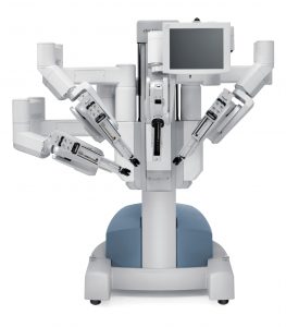
 No more ‘long and the unsightly scar on his neck after a cervical esophagus, the sign will remain’ hidden in the armpit. Italy is leading the way to surgery with the use of robots by Transaxillary. The first intervention of this kind in the world and ‘was executed on July 8 in Modena Hospital Baggiovara by Professor Gianluigi Melotti and his team consisting of Micaela Small, Barbara and John Mullineris Hills, using the Da Vinci surgical robot on a patient of 71 years. The man had a massive (about 5cm) diverticulum of the cervical esophagus (the initial part of the esophagus, located in the neck) that caused dysphagia and regurgitation. With the experience gained with the transaxillary robotic thyroidectomy (Modena and ‘one of t he major European centers for this technique, with more’ than 130 cases performed from September 2010), starting from the left armpit and creating a subcutaneous tunnel first and then through the divaricandoli neck muscles, without dissect, respecting hence the complete integrity ‘, and’ status update has reached the esophagus, a little more ‘deep thyroid in the neck. Raised the thyroid with the aid of the “Modena retractor” (a special retractor also made by the team Modena) and the Da Vinci robot, and ‘proceeded to the delicate esophagus isolation, respecting the noble structures of the neck (thyroid and parathyroid , recurrent laryngeal nerve, whose injury causes severe alterations of voice and breathing, trachea, carotid artery and jugular vein) exposure of the diverticulum (under continuous endoscopic control) and its complete removal thanks to a stapler cut and paste. The transaxillary robotic technology has made it possible to perform the surgery without the unsightly and long incision in the neck, with a quality ‘vision that greatly facilitate compliance with the precise structures described and resection of the diverticulum to his collar.
  The greater precision guaranteed by the surgical robotic technology greatly reduces the possible complications of this procedure (such as bleeding, fistulas, infections), and the wound is completely hidden under the armpit . The patient performed a radiological control in the third postoperative day, the control that has documented the success of the intervention: the disappearance of the diverticular sac with restoration of normal morphology of the esophagus and absence of esophageal fistulas. After a semi-liquid diet for about a week, the man and ‘returned to a normal diet. The cervical esophageal diverticulum Zenker’s also called the German pathologist who discovered ‘, and’ substantially un’estroflessione mucosa (a sort of bag) through an area of ??weakness in the muscle wall (Killian’s triangle). This and ‘the most’ common esophageal diverticulum and accounts for about 60-70%, even 90% for some of these diseases of the esophagus: and ‘still not considered a disease frequently encountered with an incidence ranging between 0, 01% and 0.11%, and a predominance of males in the age and ‘middle-age. The symptomatology and ‘characterized by dysphagia (difficulty’ on swallowing), typically due to the large size of the diverticulum that compresses and deflects the esophageal lumen. To this is accompanied by the regurgitation of undigested material (also related to previous meals). There may be frequent inhalations, with recurrent bronchopneumonia. Rare ly, and in extreme cases, you can ‘be gurgling in the cervical region. The traditional surgical technique involves an incision, long enough, cervical, usually on the left side of the neck, which allows the execution of a suspension (diverticulopessi) or resection (diverticulectomy) of the diverticulum. There is also the endoscopic technique, with access via endo-oral, that provides for the section of the septum between the esophagus and diverticulum or in the section / suture of the same by means of a surgical instrument, a linear stapler, introduced through the mouth. All endoscopic techniques, however, ‘compared to surgery, do not remove the diverticulum, simply increase the diameter of the collar, reducing the symptoms and possible complications, but not’ remove the diverticulum itself and are indicated only in those patients who have comorbidities ‘multiple high anesthetic risk and it is not possible to proceed with surgery.
Sunday, July 21, 2013
Health, cervical esophagus: the long scar is just a memory. Here's the ... - Weather Web
Subscribe to:
Post Comments (Atom)
No comments:
Post a Comment